Article Optical instrument human eye
Parts of the eye
The cornea is a transparent, hard membrane covering the front of the eye. The cornea refracts the beam of light entering the eye.
The pupil is black parts of the eye. The pupil is black, therefore absorb almost all the light. A pupil is a hole through which light passes into the eye. The larger the diameter of the pupil, the more light enters the eye and the smaller the size of the pupil, the less light comes to the eye.
The iris controls the size of the pupil. When the environment around the eye is very bright, the iris minimizes the pupil. Conversely, when the environment around the eyes is less bright, the iris enlarges the pupil so that the amount of light entering the eye is large. If you have observed the difference between the cat’s eye during the day and night, then you can easily understand the function of the iris.
Ciliary muscles function to change the curvature of the eye lens. Changes in the eye lens affect the focal length of the eye lens. If the eye observes objects at a great distance, the ciliary muscles relax so that the radius of the lens curvature increases and the focal length of the lens increases.
When the eye observes objects at close range, the ciliary muscles contract so that the radius of the lens curvature decreases and the focal length of the lens decreases. So, indirectly, the function of ciliary muscles is to control the focal length of the eye lens so that the eye can focus the image of the object correctly. The change in focal length of the eyepiece is called eye accommodation.
The eye lens is a convergent lens or convex lens or positive lens. Although more refraction of light occurs in the cornea, the lens of the eye also refracts and focuses the beam of light so that the image formed right on the retina. An image must be formed on the retina so that objects can be seen clearly. The retina functions to convert light waves into electrical signals, which are then passed through the optic nerve to the brain.
Accommodation by a normal eye
Eye accommodation is the adjustment of focal length (f) of the eye lens with the distance of the object from the eye so that the image can be focused on the retina. The focal length of the eye lens is the distance between the eye lens and the focal point of the lens (F). When the eye accommodates, the curvature of the eye lens changes, causing the radius of curvature of the eyepiece to change and hence the focal length of the eye lens also changes.
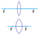 If the eye observes an object at a far point, the ciliary muscle relaxes, causing the eye lens to become flattered so that the radius of curvature and the focal length of the lens increase. The far point is the farthest distance that the eye can focus on, where the distant point of the normal eye is infinite. When the ciliary muscles relax so that the focal length of the eye lens becomes longer and can focus on the object at a distant point, the eye has minimum accommodation.
When the eye observes objects at close range, the ciliary muscles contract, causing the eye lens to become more curved so that the radius of curvature and the focal length of the lens decrease. The object distance to the nearest eye that can still focus by eye is called the near point. The average human has a near point of 25 cm. When the ciliary muscles contract so that the focal length of the lens of the eye becomes shorter and can focus on the object at a near point (25 cm), the eye has maximum accommodation.
Formation of image
The far point
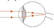 If the object is infinite, the eye lens turns flattered until the focal length (f) of the lens is equal to the image distance (di). The image distance is the distance between the lens and the point where the beam of light refracted by the eye intersects. The point where the rays of light intersect, coincide with the retina. So can be concluded that when the eye observes an infinite object, the curvature of the eye lens turns flat until the focal length of the lens (f) equals the distance between the lens and the retina. The light beam must intersect in the retina so that light can be converted into electrical signals in the retina.
The following is a mathematical proof of why when an object is at infinity, the focal length of the lens (f) is equal to the image distance (di). The relationship between focal length (f), the object distance (do) and the image distance (di) are expressed through the following equation.
1/f = 1/do + 1/di —– Equation 1
The eye lens is a converging lens. Therefore, the focal length (f) is positive. The object distance (do) is at infinite, so replace the object distance (do) in the equation 1 with an infinity symbol.
1/f = 1/~ + 1/di
1/f = 0 + 1/di
1/f = 1/di
di = f —– Equation 2
Equation 2 shows that on converging lenses if the object is at infinity, the focal length (f) is equal to the image distance (di).
The near point
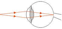 What if the object is close to the eye? Previously, it was explained that the near point of the normal eye or the distance of the nearest object that can be focused on properly by the normal eye is 25 cm. Thus, the focal length (f) of the eye lens must be smaller than the object distance (do) 25 cm. The focal length of the lens of the eye must be smaller than the distance of 25 cm so that the lens of the eye produces the real image. The eye lens is a convex lens that can form a virtual image and a real image. A real image is produced by a convex lens when the focal length (f) is smaller than the object distance (do). This has been discussed in the topic of the image formation of the convex lens.
Estimate the focal length (f) of the eye lens system:
The near point of the normal eye is 25 cm so that the distance of the nearest object (s) is 25 cm. The distance between the cornea and the retina is about 2.5 cm. The image is clear if the beam of light falls on the retina so that the distance between the cornea and retina is considered as the image distance (di). So, the image distance (di) is 2.5 cm.
1/f = 1/do + 1/di
1/f = 1/ 25 + 1/ 2.5 = 1/25 + 10/25 = 11/25
f = 25/11
f = 2.27 cm
Based on the results of the above calculations, it can be concluded that the focal length (f) is smaller than the image distance (di). The difference between the image distance (di) and the focal length (f) is 2.5 cm – 2.27 cm = 0.23 cm. The focal point of the cornea-eye lens system is 0.23 cm to the left of the image or retina.
The cornea-eye lens system is a convex lens so that the image is formed on the retina even though it is real but inverted. Even though the image on the retina is inverted, the brain’s nerve turns it upright.

